Silent Suffocation: Deconstructing Hypoxia – From Cellular Distress to Catastrophic Organ Failure

Section One: Physiological Definition and Cellular Basis (The Silent Life Crisis)
Hypoxia is a critical medical condition defined by an inadequate oxygen supply to body tissues, insufficient to meet their metabolic needs. This oxygen deficit disrupts cellular energy production, primarily affecting mitochondrial oxidative phosphorylation, which generates most cellular ATP. It is essential to distinguish hypoxia from hypoxemia: hypoxemia refers specifically to low oxygen levels in arterial blood, whereas hypoxia denotes insufficient oxygen at the tissue level. For example, in carbon monoxide poisoning, arterial oxygen may appear normal, but tissue oxygen delivery is impaired due to hemoglobin binding interference.
Section Two: The Molecular Blueprint: Cellular Response Under Stress (The HIF-1 Factor)
Cells respond to oxygen deprivation by activating Hypoxia-Inducible Factor 1 (HIF-1), a transcription factor that senses oxygen levels. Under normal oxygen conditions, the HIF-1α subunit is degraded rapidly; hypoxia inhibits this degradation, allowing HIF-1α to accumulate, dimerize with HIF-1β, and activate genes that promote survival. HIF-1 induces a metabolic shift from aerobic respiration to anaerobic glycolysis, increasing lactic acid production and causing metabolic acidosis. It also stimulates erythropoietin production to increase red blood cell synthesis and promotes angiogenesis to improve tissue oxygenation. These adaptations are crucial for short- and long-term survival under low oxygen conditions and represent potential therapeutic targets.
Section Three: Causes and Clinical Patterns (Crisis Classification)
Hypoxia is classified into four main types based on the defect in oxygen delivery or utilization:
- Hypoxic (Respiratory) Hypoxia: Caused by impaired pulmonary gas exchange or ventilation, seen in COPD, asthma, pneumonia, pulmonary edema, and high-altitude exposure.
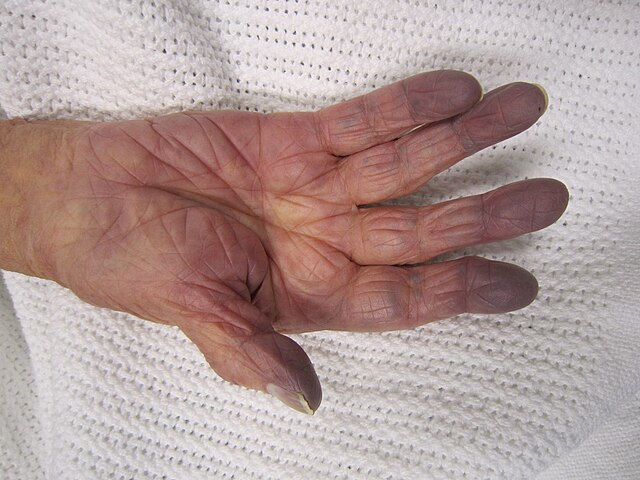
- Anemic Hypoxia: Due to reduced oxygen-carrying capacity of blood, as in anemia or carbon monoxide poisoning, where CO binds hemoglobin with high affinity, preventing oxygen transport.
- Stagnant (Circulatory) Hypoxia: Results from inadequate blood flow despite normal oxygen content, common in heart failure, shock, and ischemia.
- Histotoxic Hypoxia: Occurs when cells cannot utilize oxygen despite adequate delivery, exemplified by cyanide poisoning which inhibits mitochondrial electron transport.
Chronic diseases often involve overlapping hypoxia types, complicating diagnosis and treatment.
Section Four: Symptoms and Clinical Warnings (Acute and Chronic Response)
The body initially compensates for hypoxia by increasing respiratory rate (tachypnea) and heart rate (tachycardia) to enhance oxygen delivery. Cyanosis, a bluish discoloration of skin, is a common sign, though absent in some types like CO poisoning. Neurological symptoms such as headache, dizziness, confusion, and impaired judgment appear early due to the brain’s sensitivity to oxygen deprivation. Severe hypoxia leads to bradycardia, coma, and irreversible brain damage. Peripheral hypoxia can cause gangrene and nerve injury. Chronic hypoxia may be masked by partial compensation but leads to systemic complications including pulmonary hypertension, right heart failure, and chronic kidney disease.
Section Five: Catastrophic Complications and Systemic Organ Failure
Prolonged hypoxia causes irreversible damage, especially in high-demand organs:
- Cerebral Hypoxia: Leads to neuronal death, cognitive impairment, coma, and permanent neurological deficits.
- Cardiovascular Failure: Chronic hypoxia induces pulmonary vasoconstriction, causing pulmonary hypertension and right-sided heart failure (cor pulmonale).
- Renal Impairment: Hypoxia worsens chronic kidney disease by reducing erythropoietin production, exacerbating anemia.
- Peripheral Tissue Damage: Acute ischemia can cause gangrene requiring urgent intervention.
Section Six: Diagnosis and Advanced Clinical Management
Diagnosis begins with pulse oximetry and is confirmed by arterial blood gas analysis measuring oxygen partial pressure and lactate levels. Imaging may identify underlying causes like pulmonary edema. Treatment focuses on restoring oxygen delivery:
- Oxygen Therapy: Supplemental oxygen via mask or nasal cannula; mechanical ventilation in severe cases.
- Hyperbaric Oxygen Therapy (HBOT): Especially effective in carbon monoxide poisoning, increasing plasma oxygen and accelerating CO removal.
- Surgical and Rehabilitation Interventions: Lung transplantation for end-stage pulmonary disease; physical therapy for neurological recovery.
Tailored treatment depends on the hypoxia type and underlying pathology.
Section Seven: Conclusion and Recommendations (Protecting the Life Stock)
Hypoxia is a complex condition beginning at the molecular level with HIF-1 activation and culminating in systemic organ failure if untreated. Early recognition of neurological symptoms and precise physiological diagnosis are critical. Preventive measures, such as carbon monoxide detector use, and comprehensive management of chronic diseases are essential. Survivors benefit from integrated rehabilitation to improve quality of life.
